 |
|||||||
|
|||||||
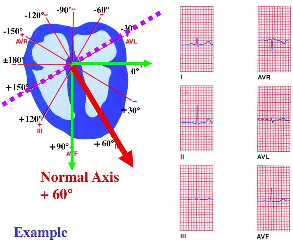
| Class, Below is an example how to determine the QRS axis and degrees | |||||||
 |
|||||||
| STEP 1: Is the QRS in Lead I mostly positive or negative? POSITIVE | |||||||
| STEP 2: Is the QRS in AVF mostly positive or negative? POSITIVE | |||||||
| STEP 3: Which lead is the biphasic lead closest to isoelectric. AVL | |||||||
| REMINDER: THE MEAN QRS AXIS IS ALWAYS PERPENTICULAR TO THE BIPHASIC LEAD. AND, STEP 1 AND STEP 2 HAVE IDENTIFIED THE QUADRANT OF THE MEAN QRS | |||||||
| STEP 4: Determine QRS Axis and degrees? Normal Axis +60 Degrees | |||||||
Mean QRS Axis Classification |
|||||||
 |
|||||||
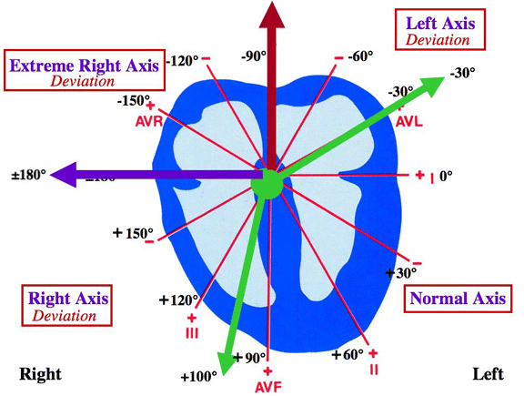
| Normal Axis: from -30 degrees to +100 degrees | |||||||
| Right Axis Deviation: from +100 degrees to ±180 degrees | |||||||
| Extreme Right Axis Deviation: from ±180 degrees to -90 degrees | |||||||
| Left Axis Deviation: from -90 degrees to -30 degrees | |||||||
| Clinical Notes on QRS Axis (Just for informational purposes). | |||||||
| The mean QRS axis represents the average of the instantaneous electrical vectors generated during the sequence of ventricular depolarization, as measured in the frontal plane. It tells us the direction the depolarization is headed in the ventricles. The normal axis ranges from -30 degrees to +100 degrees although some sources use the -30 to +90 degrees range. Right axis deviation is seen on the ECG when more electrical forces are moving to the right than normal. This is usually due to hypertrophy of the right ventricle (RVH). Causes of right axis deviation include COPD, pulmonary emboli, valvular disease, septal defects, and pulmonary hypertension. An axis of +90 is common in persons with emphysema. This so-called “vertical heart” reflects both the rotation of the heart downward as the diaphragm position drops due to air trapping, and some degree of hypertrophy of the right ventricle. Left axis deviation occurs when additional electrical forces move to the left (hypertrophy), or when the time required for the electrical activity to move over the ventricle is prolonged (LBBB, left ventricular dilation). Causes of left axis deviation include hypertension, aortic stenosis, subaortic stenosis, mitral regurgitation, and left ventricular conduction defects. |
|||||||
|