| February 1 to February 5: Motor Unit Recruitment, Skeletal Muscle Contraction Mechanisms OBJECTIVES: Be able to explain the recruitment of Type I, Type IIa and Type IIx motor units Be able to explain in detail the mechanisms of a muscle contraction (action) Be able to explain the physiological steps of the arrival of a nerve impulse at the neuromuscular junction Be able to explain the physiological commonality of the sarcolemma, motor end plate and transverse tubules Be able to define several terms related to the physiology of muscle contraction Please note there are THREE YOUTUBE videos with this week's lessons. Click Here to Download Exam 1 Part C due Friday February 5 by 12 Midnight Let's start this section by watching this Youtube Video on motor unit recuitment in skeletal muscle Click here for the Motor Unit Recruitment YOUTUBE video Let's Review, from the YOUTIBE Video, a few concepts: With motor unit recuritment, are specific motor units 'isolated' or 'emphasized'? (emphasized) Compare the diameter of the nerve and diameter of the muscle fiber of a Type I and Type IIa motor unit. (the diameter is bigger in the Type IIa motor unit) Compare the diameter of the nerve and diameter of the muscle fiber of a Type IIa and Type IIx motor unit. (the diameter is bigger in the Type IIx motor unit) |
||||||||
| Motor Unit Recruitment | ||||||||
 |
||||||||
| Learning Tip: Notice in the above graphic how the author is depicting the motor unit with ONE nerve innervating multiple muscle fibers on the left side of the graphic. With the top picture, the load is light for the exerciser and the Type I motor units have been innervated. In the middle picture there are more plates on the bar and thus more force to overcome. The Type IIa is doing most of the work with the help of the Type I. In the bottom picture, there are many plates on the bar requiring near maximal exertion of the exercise. This recruits the most powerful motor units in the body, the Type IIx and you see the Type IIa and Type I are helping. Please note with any load of work you notice how the Type I fibers are always firing. |
||||||||
|
|
||||||||
| The Mechanisms of Muscle Contraction (Part 1) | ||||||||
| Click here to watch the Mechanisms of Muscle Contraction Part 1 YOUTUBE video |
||||||||
 |
||||||||
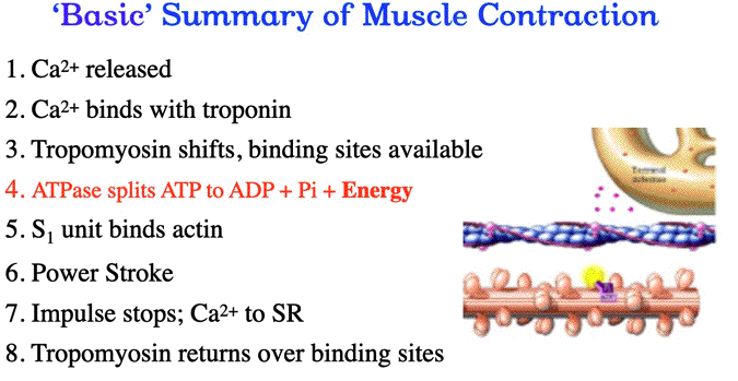
| Here are some questions from this YOUTUBE video lecture (below) | ||||||||
| Where is the calcium ion housed in muscle? (sarcoplasmic reticulm) Calcium has a strong affinity to what protein? (troponin) Which protein shifts during a muscle action? (tropomyosin) What causes the S1 units to disconnect from actin binding sites? (fresh ATP) Which protein on actin is composed of 'three component structures'? (troponin) The binding sites on actin are covered by what protein? (tropomyosin) After the nerve impulse stops what happens to the calcium ions? (pumped back into the sarcoplasmic reticulum) The movement of actin over myosin is called a ___________? (power stroke) Before myosin S1 units attach to binding sites on actin, what has to happen to ATP? (Splits into ADP+ Pi+ energy) |
||||||||
| The Mechanisms of Muscle Contraction continued | ||||||||
| Click here to watch the Mechanisms of Muscle Contraction Part 2 YOUTUBE video |
||||||||
 |
||||||||
|
||||||||
|