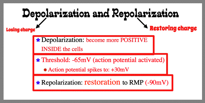
| August 17 - 21: Review Heart Anatomy, EKG Intro, Review of Cardiovascular Physiology and Anatomy OBJECTIVES: Be able to identify key anatomical structures in the heart Be able to discuss the blood flow through the systemic and pulmonary circuits Be able to explain the cardiac cycle in detail Be able to name the two major cell system of a working heart Be able to name some key pioneers of EKG Be able to draw and label the cadiac conduction system of the heart Be able to explain depolarization, repolarization, and resting membrane potential (RMP) Be able to describe the electrical charges in heart at RMP, threshold and depolarization Click Here to Download Exam 1 Part A due Friday August 21 by 12 Midnight Note to Class: There are 3 Youtube Videos with this Week (see below): Intro to EKG, First Lecture Video, and the Cardiac Conduction System Video Please watch the introductory Youtube video to EKG (IF YOU HAVEN'T VIEWED YET). Dr. Kravitz discusses how the class will be taught this semester and his academic goals and expectations for all students in PEP326L. He introduces the EKG Mantra: Study/Learn/Know. Click here for the Introduction To EKG YOUTUBE video We are ready for our first EKG YOUTUBE video which Reviews Heart Anatomy and Cardiovascular Physiology. Click here to get started on our First 20-minute EKG YOUTUBE Lecture |
||||||||
| Heart Anatomy Review | ||||||||
 |
||||||||
 |
||||||||
 |
||||||||
| With the two pump 'schematic' of the heart in the YOUTUBE video it was clear that the four chambers of the heart actually function similar to two pumps. The right side of the heart pumps to the lungs (pulmonary circuit) and the left side of the heart pumps to the periphery of the body (systemic circuit). | ||||||||
| Blood Flow Through Pulmonary and Systemic Circuit | ||||||||
 |
||||||||
| Review: What atrioventricular (AV) valve is on the right side of the heart? (Tricuspid). What atrioventricular (AV) valve is on the left side of the heart? (Mitral). Name the semilunar valves. (Aortic semilunar and pulmonary semilunar valves). |
||||||||
| Cardiac Cycle: Three Phases | ||||||||
 |
||||||||
| What are the TWO major cell systems of a working heart? Electrical (autorhymic cells, pacemake cers) and Mechanical (contractile cells, myocardial cells) The electrocardiogram depicts the electrical activity of the heart. |
||||||||
| Cardiac Conduction EKG YOUTUBE Video below: | ||||||||
| Click here to watch the EKG YOUTUBE Lecture on the Cardiac Conduction System |
||||||||
| Cardiac Conduction System of the Heart | ||||||||
 |
||||||||
| Depolartization begins at SA node, then travels to Bachmann's Bundle, AV node, bundle of His, Right and Left Bundle Branches (left anterior and posterior fascicle) and lastly to Purkinje fibers. Class, please note that Bachmann's bundle is an interatrial conduction pathway (which means it conducts the depolarization from the SA node to the left atria). |
||||||||
 |
||||||||
 |
||||||||
| Introducing Some Pioneers of EKG | ||||||||
| What pioneer published the 1st electrical recording of the human heart. Augustus Waller | ||||||||
| What pioneer discovered blood flow through the human body? William Harvey | ||||||||
| What pioneer published the first EKG? William Einthoven | ||||||||
| What pioneer discovered the interatrial conduction pathway (between right and left atria)? Jean George Bachman | ||||||||
| What pioneer discovered the purkinje fibers? Johannes Evangelista Purkinje | ||||||||
| What pioneer discovered the bundle of His? Wilhelm His Jr | ||||||||
| Exam 1 Part A: Click here to get questions: Exam 1 Part A is due by 12midnight on August 21. No late papers accepted. |
||||||||
|